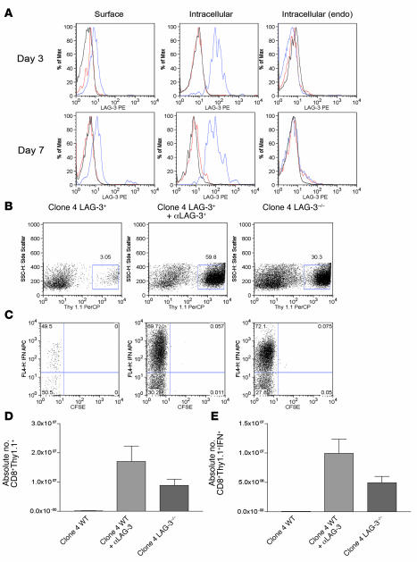
Figure 1. LAG-3 blockade enhances the accumulation of HA-specific CD8+ cells in a model of self tolerance.
We transferred 106 LAG-3+/+ or LAG-3–/–CD8+Thy1.1+ clone 4 CD8+ cells into Thy1.2+C3-HAhigh mice. Mice receiving antibody were given 0.2 mg αLAG-3 i.p. at the time of transfer and 3 days later. At indicated time points after transfer, single-cell suspensions were made from lungs and analyzed for LAG-3 expression and function. (A) CD8+ and Thy1.1+ were gated to determine expression of LAG-3 on CD8+ T cells taken directly from lungs of C3-HA mice. Both surface expression (left) and intracellular staining (middle and right) were performed for LAG-3 expression. Endogenous (endo) CD8 populations within lungs were also assessed. Blue lines, LAG-3; red lines, rat IgG1 isotype; black lines, LAG-3–/– cells. Max, maximum. (B) Lungs from C3-HA mice were analyzed 7 days after transfer for percentage of CD8+Thy1.1+ cells by staining for Thy1.1 (C). Clonotypic cells from B were analyzed for division by dilution of CFSE and IFN-γ production by intracellular staining after stimulation in vitro in the presence of HA peptide plus monensin for 5 hours. SSC-H, side scatter. (D and E) Absolute numbers of clonotypic and IFN-γ+ clonotypic cells/lung after αLAG-3 treatment or transfer of LAG-3–/– clone 4 cells. The mean from 3 mice per group is shown. Each experiment was performed 3 times with data from 1 representative experiment shown.

