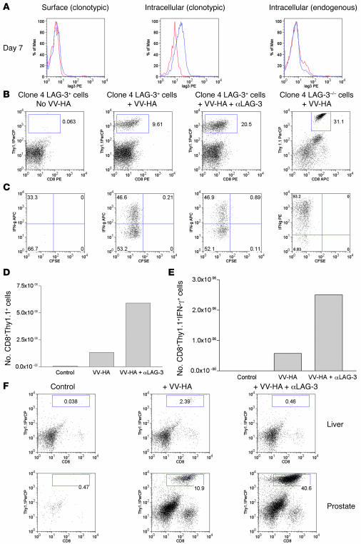
Figure 2. LAG-3 blockade enhances the accumulation of clone 4 CD8+ effectors within HA-producing prostates of ProHA × TRAMP mice.
(A) In vivo expression of LAG-3 on clonotypic CD8+ T cells. We transferred 2 × 10⁶ Thy1.1+ clone 4 CD8+ cells into VV-HA–injected ProHA × TRAMP mice. Seven days later, prostates were harvested and single-cell suspensions were gated on CD8+ and Thy1.1+ cells and analyzed for expression of LAG-3. Both surface (left) and intracellular staining (middle and right) were performed for detection of LAG-3. Blue line, LAG-3; red line, rat IgG1 isotype; black line, LAG-3–/– cells. (B) αLAG-3 enhances the accumulation of clonotypic cells in prostates. We transferred 106 LAG-3+/+ or LAG-3–/–CD8+Thy1.1+ clone 4 CD8+ cells into ProHA × TRAMP mice. Mice were given VV-HA, VV-HA plus αLAG-3, or nothing at the time of transfer. Mice receiving αLAG-3 were given another dose 3 days later. Seven days after transfer, prostates and livers were collected and homogenized, and single-cell suspensions were analyzed for IFN-γ by intracellular staining after stimulation in vitro in the presence of HA peptide plus monensin for 5 hours. Prostates were analyzed for (B) percentage of CD8+Thy1.1+ and (C) percentage of IFN-γ+ clonotypic cells. Absolute numbers of pooled prostates to determine (D) prostate-derived clonotypic cells and (E) IFN-γ+ clonotypic cells are shown. Error bars are absent because pooling was necessary to count CD8+ cells from prostate tissue. The results are representative of at least 4 experiments. (F) Enhanced accumulation of clone 4 CD8+ cells using αLAG-3 is specific for organs expressing cognate antigen. Livers from ProHA × TRAMP mice do not express the HA antigen (data not shown) and fail to attract and promote division of HA-specific CD8+ T cells. Data from 1 of at least 3 individual experiments are shown. PerCP, peridinin chlorophyll protein.

