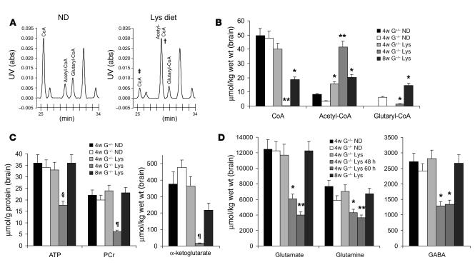
Figure 5. Age-dependent brain biochemical changes with lysine diet exposure.
(A) Chromatogram of acyl-CoA esters from brain extract of weanling Gcdh–/– mice on normal or lysine diet shows large accumulation of acetyl-CoA (†) and depletion of free CoA (‡) after 48 hours of lysine diet. Abs, absorbance. (B) Free CoA, acetyl-CoA, and glutaryl-CoA changes in brain extract of weanling and adult Gcdh–/– mice and heterozygous controls on normal or lysine diets. Mean ± SEM, *P < 0.01; **P < 0.001. n = 6 each group. (C) ATP, phosphocreatine (PCr), and α-ketoglutarate changes in cortex of weanling and adult Gcdh–/– mice and heterozygous controls on the lysine diet compared with normal diet. Mean ± SEM, ΧP < 0.04; ζP < 0.02. n = 4 each group. (D) Glutamate, glutamine, and GABA levels in weanling and adult Gcdh–/– mice and heterozygous controls on the lysine diet compared with normal diet. Mean ± SEM. n = 6 each group.

