
Figure 4. Localization of SPIN90 and palladin in PDGF treated U251 cells.
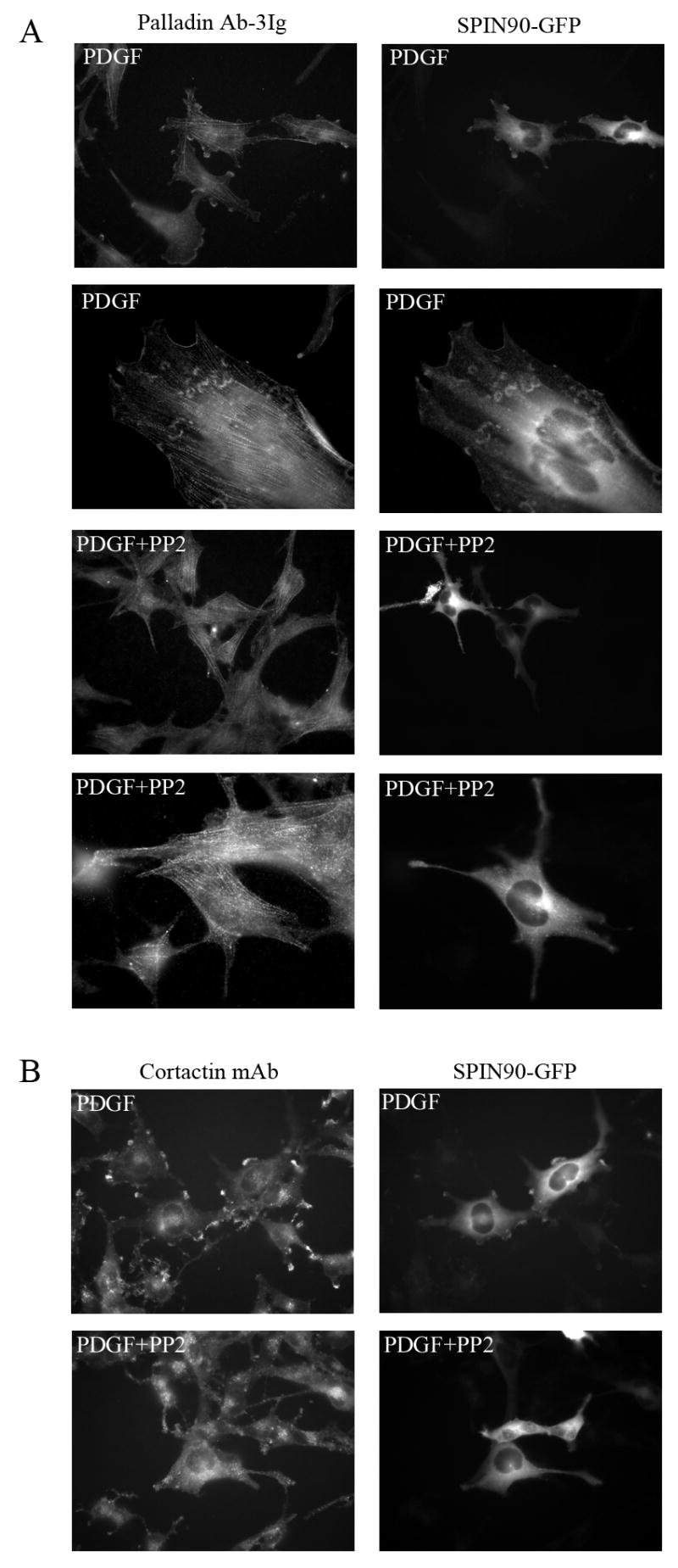
(A) U251 cells transfected with GFP-SPIN90 construct were serum-starved for 16h and stimulated with PDGF for 10 minutes. Cells were fixed and stained for endogenous palladin. Both palladin and SPIN90 are relocated to the PDGF-induced membrane ruffles (top row) and actin containing wave-like structures (second row). Treatment with the Src kinase family inhibitor PP2 prior to PDGF prevents the redistribution and both proteins remain in their normal subcellular positions (third and fourth row). (B) In the same experiment, the co-localization of cortactin, a known Src kinase substrate, and SPIN90 was compared. Cortactin is known to re-locate to the membrane after PDGF treatment and, as expected, cortactin co-localized with SPIN90 in the membrane ruffles in PDGF-treated cells. This re-location was Src-dependent as it was abolished by PP2.