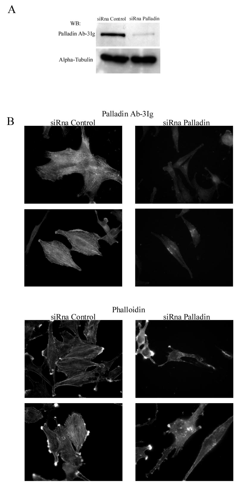
Figure 6. Depletion of palladin results in disruption of actin cytoskeleton and loss of stress fibers.

(A). U251 cells were transfected with a palladin specific siRNA oligonucleotide or with a non-targeting control siRNA. The specific siRNA markedly reduced the amount of palladin as compared to control. The alpha-tubulin blot serves as a loading control. (B). Transfected cells were stained with Ab-3Ig palladin antibody and with labeled phalloidin to visualize F-actin. The intensity of palladin staining is markedly reduced and the remaining staining pattern was diffuse compared to the control cells, which showed the typical stress fiber dense region staining. Depletion of palladin resulted in loss of stress fibers and disorganization of the cytoskeleton as visualized by phalloidin staining.
