
Figure 1.
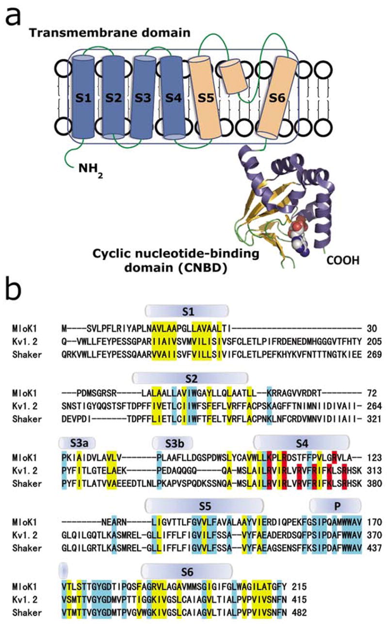
(a) Proposed topology of one MloK1 subunit. Each subunit consists of an N-terminal transmembrane domain and a C-terminal cyclic nucleotide-binding domain (CNBD). Helices S1-S4 (blue) are the putative voltage sensors, and helices S5, S6, and the pore helix (light orange) form the pore region. CNBD is shown in ribbon representation, bound cAMP is shown space-filled in cpk. (b) Sequence alignment of the transmembrane domains of MloK1 (Mesorhizobium loti), Kv1.2 (Rattus norvegicus) and Shaker (Drosophila melanogaster). The secondary structures of Kv1.2 are indicated. Residues of similar chemical characteristics are yellow, identical residues are light blue, and positively charged amino acids (arginine, lysine) in S4 are red.