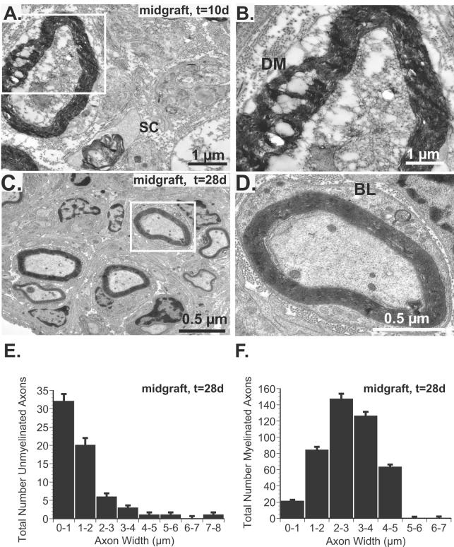
Fig 5.
A. Electron micrograph of central graft cross-section 10 days after surgery confirms the absence of viable axons although SCs (SC), migrating into the acellular allografts are noted. B. A magnified view of the inset from panel A. reveals degenerated myelin (DM) and no viable axons. C. By 28 days, both myelinated and unmyelinated axons are noted. D. By 28 days, however, intact, myelinated axons are noted and an intact basal lamina (BL), characterized by double basement membranes are seen in this magnified inset from panel C.. E. Quantification and stratification of unmyelinated axons according to width reveals a strong bias towards small diameter axons (82% ≤ 2 μm). F. Similarly, only 1% of axons were >5 μm in diameter 28 days after surgery.

