
Figure 4.
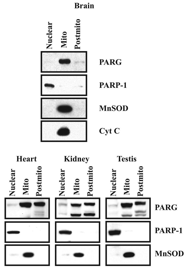
Subcellular localization of PARG. 20 μg of protein of each subcellular fraction from the brain, heart, kidney and testis were separated on SDS-PAGE and transferred to a nitrocellulose membrane. The membrane was blotted with α–PARP-1, α–PARG, α–MnSOD and α–Cyt C. Western blots were developed by chemiluminescence using SuperSignal. These results were replicated three times with similar results.