
Figure 5.
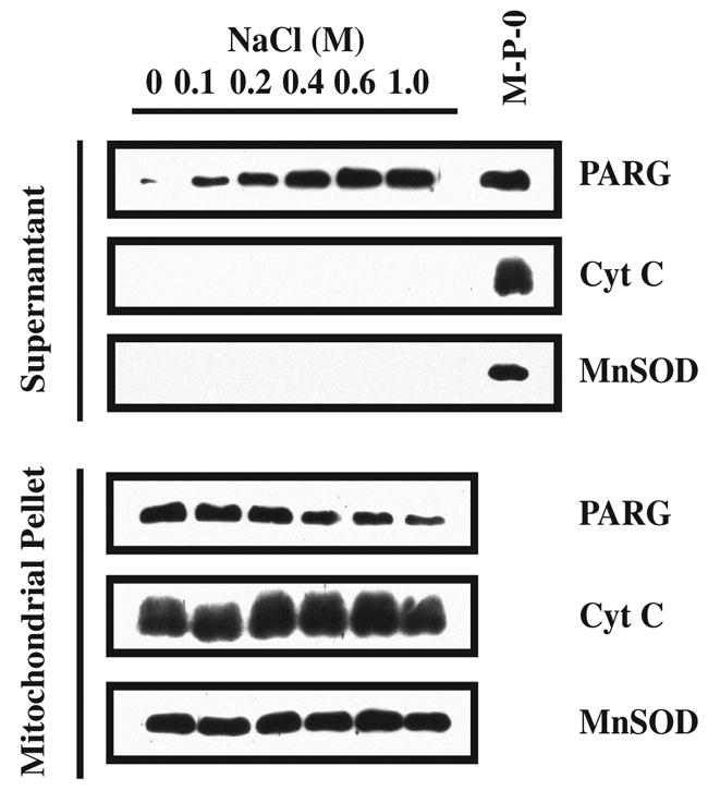
Mitochondrial PARG salting out. The mitochondrial pellet (P2) was aliquoted in separate tubes and centrifuged. The individual pellets were then resuspended in 200 μl of subfractionation buffer containing increasing concentrations of NaCl. The mitochondrial suspensions were centrifuged 30 min at 10,000 x g. The proteins contained in the supernatants and pellets were resolved on SDS/PAGE gel, subjected to Western blot analysis and blotted with α–PARG, α–MnSOD and α–Cyt C. Western blots were developed by chemiluminescence using SuperSignal. These results were replicated two times with similar results. M-P-0: mitochondrial pellet at 0 M NaCl.