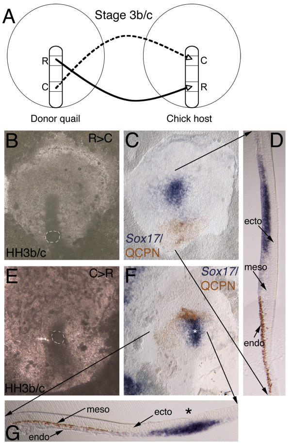
Figure 3.
Streak to streak quail-chick chimera transplants. (B, C, E, F) Whole mount ISH, anterior to the top (Sox17, blue) and immunochemistry with anti-QCPN antibody (brown). (D, G) 50 μm gelatin sections. (A) Schematic depicting the experimental manipulation. Donor quail stage 3b/c with either rostral to caudal streak transplant (C-D) into chick host at same stage or caudal to rostral transplant (E-G). A second population of rostral streak cells, called "tip" cells (not diagrammed; see text and Figure 4) was also transplanted more caudally, and, conversely, caudal streak cells were transplanted to the tip site. (B) Host embryo at stage 3b/c within 1 hour of transplanting quail tissue from rostral to caudal streak, with fully integrated site highlighted by white dashed circle. (C) Same embryo after 4–6 hours incubation fixed and stained with Sox17 and anti-QCPN. (C, D) Sox17 is expressed in streak and more lateral migrating endoderm, but down regulated in transplanted cells, which are migrating normally. Sagittal section shows transplanted cells still retain endoderm germ layer position. (E) Caudal transplant integrated (white dashed circle) within rostral streak of stage 3b/c embryo and (F) after 6–4 hours incubation. Cells migrate normally away from streak. (G) Transverse section shows Sox17 in streak (asterisk), but absent from transplanted quail cells that nonetheless maintain mesoderm germ layer position.

