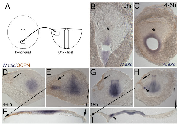
Figure 7.
Streak to CBI quail-chick chimera transplants. (B-I) ISH (Wnt8c, blue) and (D-I) immunochemistry (anti-QCPN antibody, brown). Anterior to top. (F, I) Black lines from E and H mark level of 50 μm transverse gelatin sections. (A) Schematic of experiment showing caudal streak isolate transplanted lateral to the streak in the area pellucida. (B) Whole mount Wnt8c ISH of quail donor following removal of the explant, showing that donor tissue is Wnt8c positive. (C) Whole mount donor quail after 4–6 hours of incubation showing that area around explant remains Wnt8c positive. (D-I) In no case does explanted tissue express Wnt8c after 4–6 hours or overnight incubation. (D-F) 4–6 hours of incubation, quail graft integrated and spreading (arrowed). (D) Explanted quail streak cells with no ectopic Wnt8c (E, F) Ectopic Wnt8c induced in overlying ectoderm reminiscent of an ectopic streak transverse to normal orientation. (G-I) Caudal streak explant into lateral area pellucida of caudal blastoderm isolate. Overnight incubation with quail cells now restricted to edge of CBI (arrowed). (G-I) Following overnight incubation no quail cells express Wnt8c. (H, I) In only in 2 cases is a small spot of ectopic Wnt8c still detectable in ectoderm (arrowhead).

