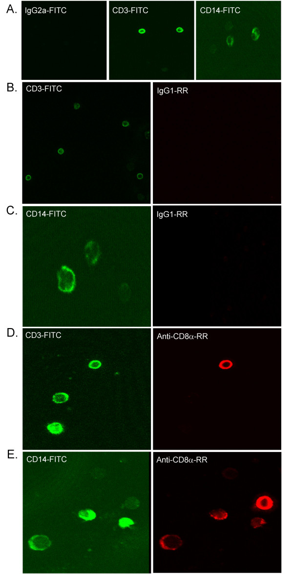
Figure 3.
CD8α is detected by confocal microscopy on peripheral blood monocytes and lymphocytes with several anti-CD8α mAb. A, CD3-FITC and CD14-FITC binding to PBMC (Green). B-E, Anti-CD8α mAb (D, E) binding to monocytes and lymphocytes in comparison to isotype mAb (B, C) (Red). Results are representative of other anti-CD8α mAb (OKT8, 51.1, 32-M4, Nu-Ts/c, and B9.11).

