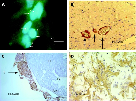
Figure 2 (A) At 3 weeks after injection, human embryoid bodies (hEBs) in the scar tissue are identified by positive FISH green signals for the X chromosome probes (arrows). The hEBs were injected 7 days after myocardial infarction. Original magnification ×1000; scale bar 10 µm. (B) Microscopic examination of normal heart treated with hEBs. Immunostaining for HLA‐ABC identified positive staining at the vessel walls (arrows), suggesting that the donor cells gave rise to new endothelial and smooth muscle cells (×400). Scale bar 20 µm. (C) Photograph of heart at week 3 after implantation of alginate scaffold (S) on infarcted heart and 2 weeks after human embryonic stem cell transfer. Immunostaining for HLA‐ABC identified viable graft that stained positive brown for the human cells (arrow), whereas rat myocardium (H) was stained negative. Scale bar = 500 µm. (D) Higher magnification of the biograft immunostained with anti‐actinin antibodies. The engrafted hEBs within scaffold remnant on infarcted heart stained positively (brown colour) for the myogenic protein, but appeared immature. Scale bar 20 µm.

An official website of the United States government
Here's how you know
Official websites use .gov
A
.gov website belongs to an official
government organization in the United States.
Secure .gov websites use HTTPS
A lock (
) or https:// means you've safely
connected to the .gov website. Share sensitive
information only on official, secure websites.
