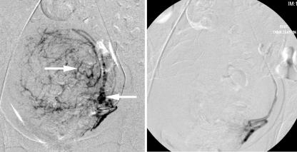
Fig 2 Left: pre-embolisation angiography performed from the right groin via a selective catheter in the left uterine artery shows the typical vascular appearances of the hypertrophied arteries supplying the uterine fibroid (arrows). Right: angiography after embolisation with polyvinyl alcohol shows contrast stasis, with no distal flow to the fibroid

An official website of the United States government
Here's how you know
Official websites use .gov
A
.gov website belongs to an official
government organization in the United States.
Secure .gov websites use HTTPS
A lock (
) or https:// means you've safely
connected to the .gov website. Share sensitive
information only on official, secure websites.
