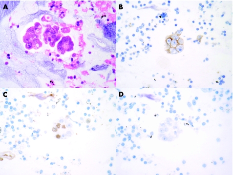
Figure 3 Photomicrographs of specimens obtained from an endobronchial ultrasound fine‐needle aspiration from a woman with a history of breast cancer and a right hilar/paratracheal mass. Groups of malignant glandular cells consistent with origin from an adenocarcinoma were identified both in the Papanicolaou‐stained ThipPrep preparation and in the H&E‐stained sections from the cell block (A). Immunistochemistry demonstrated that the tumour cells expressed BerEp4 (B) and showed focal nuclear staining with thyroid transcription factor1 (C). No staining with antibodies to oestrogen receptor was identified (D). A subsequent mediastinoscopy confirmed the presence of adenocarcinoma, which was morphologically different from the breast lesion and consistent with origin from a bronchial carcinoma, (original magnification ×400).

An official website of the United States government
Here's how you know
Official websites use .gov
A
.gov website belongs to an official
government organization in the United States.
Secure .gov websites use HTTPS
A lock (
) or https:// means you've safely
connected to the .gov website. Share sensitive
information only on official, secure websites.
