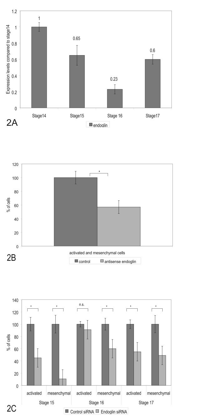
Figure 2.
Endoglin expression changes during development but is required for EMT. (A). Levels of endoglin mRNA expression in AV canal segments measured by Real Time PCR. Dissected AV canal segments were collected for mRNA analysis. Endoglin expression levels decreased at stage 16 and increased at stage 17 when mesenchymal cells begin to invade the cushions. Data represent duplicate experiments with three repetitive reactions per stage. Data was normalized to total cDNA in each reaction. Changes are graphed relative to stage 14 and relative values are displayed above each bar. (B). Cells from antisense endoglin treated explants (60 pooled explants, stages 15 -17 at time of collection) showed a loss of mesenchymal and activated cells (40% reduction) after 24 hrs compared to control oligonucleotides. (C) Staged explants were specifically examined for activation and invasion after siRNA endoglin treatment. Loss of endoglin produced the greatest loss of mesenchyme in stage 15 explants but a loss was observed in explants collected from each of the other stages. Activation of endothelial cells on the surface was unaffected in stage 16 explants and significant in both the younger and older explants. siRNA effects on mesenchymal and activated cells are compared to effects of control siRNA. Activated cells are defined as cells without attached neighboring cells remaining on the surface of the gels. A minimum of 15 explants were counted for each siRNA experiment. Error bars indicate the SEM. Significant p values, smaller than 0.05, are indicated by an asterisk (*) in each panel.

