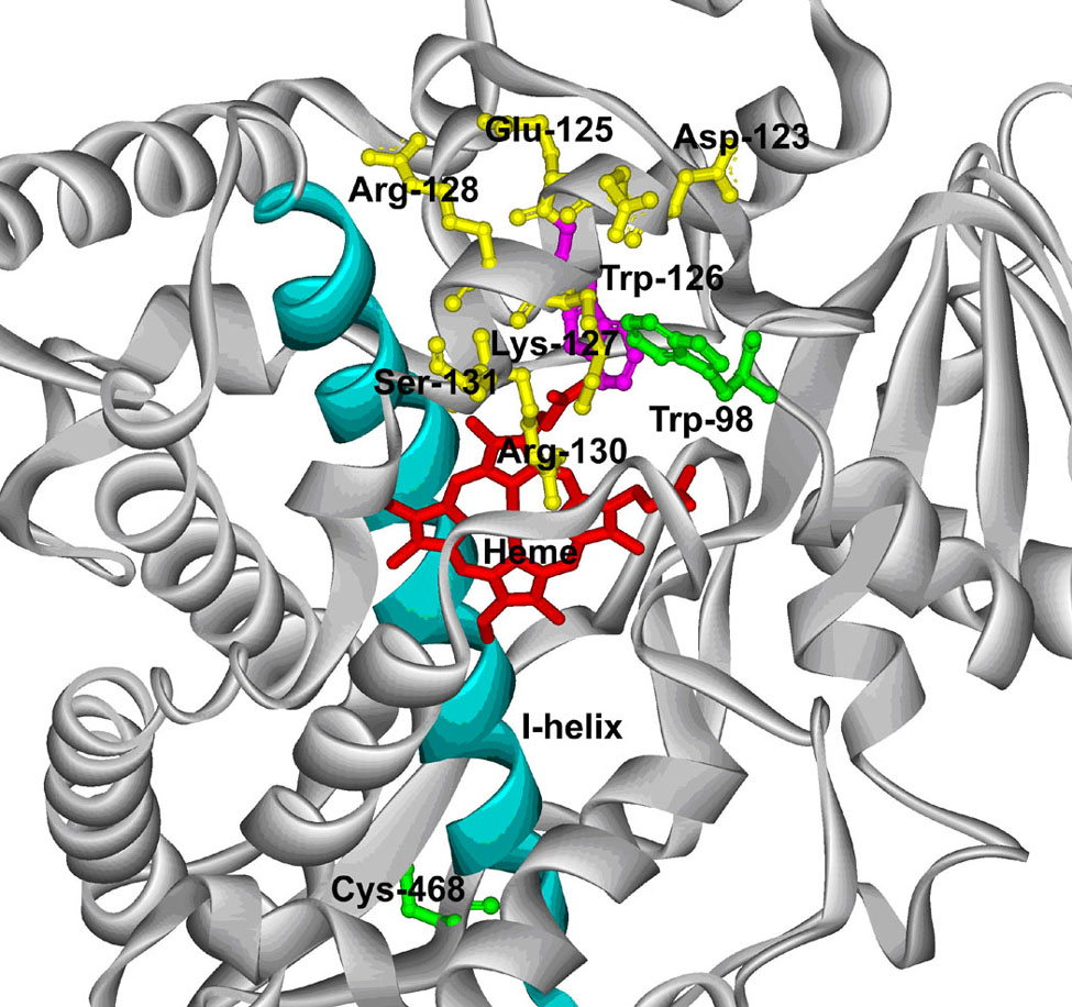
Figure 8.
A partial structure of the CYP3A4 C98W mutant. The Cys-98 of the X-ray crystal structure of CYP3A4 (PDB code: 1W0E) was substituted by a tryptophan residue using the mutate feature of DeepView 3.7 as described in Experimental Procedures. The substituted Trp-98 (green) is shown in a ball and stick model with respect to Trp-126 (purple), Cys-468 (green) and other residues (yellow) in the C-helix. The I-helix (cyan) is also shown with respect to a stick model of the heme (red).

