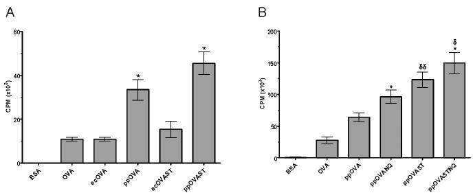
Figure 1. Effect of mannosylation on CD8+ MHC I-restricted lymphoproliferation.
BMDCs were cocultured with CD8-purified T cells from OT-I mice in the presence of the indicated antigens for 72 h before pulsing with [3H]-thymidine for 18 h. A. Comparison of E. coli and P. pastoris-derived antigens. Data are the mean ± SEM (n=3) of a representative of three independent experiments. *, p<0.01 (comparing ppOVA with ecOVA and ppOVAST with ecOVAST). B, Comparison of antigens with varying degrees of N- and O-linked mannosylation. Data are the mean ± SEM (n=15) of five independent experiments, each of which was performed in triplicate. *, p<0.01 (comparing ppOVANQ with ppOVA and ppOVASTNQ with ppOVAST). δ, p<0.05 (comparing ppOVASTNQ with ppOVANQ). δδ, p<0.01 (comparing ppOVAST with ppOVA).

