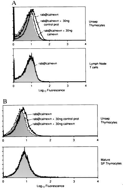
Figure 3.
Calnexin’s expression on the cell surface is developmentally regulated. (A) Single-cell suspensions from fresh C57BL/6 mouse thymic and lymph node explants were either stained directly (Unsep. Thymocytes) or after depletion of non-T cells (Lymph Node T cells). (B) Single-cell suspensions of thymocytes were stained before (Unsep. Thymocytes) or after immature cells were eliminated using anti-HSA and complement (Mature SP Thymocytes). Cell suspensions were stained with a rabbit polyclonal serum directed at calnexin’s lumenal domain (rab@calnexin, solid lines) and bound Ab were visualized with fluoresceinated-goat anti-rabbit IgG. Specificity of the anti-calnexin staining was demonstrated by preincubating the anti-calnexin Ab with 30 ng of control bacterial extract (short dashes) or extract containing calnexin fusion protein (long dashes). Cells stained with preimmune serum are indicated by shaded curves.

