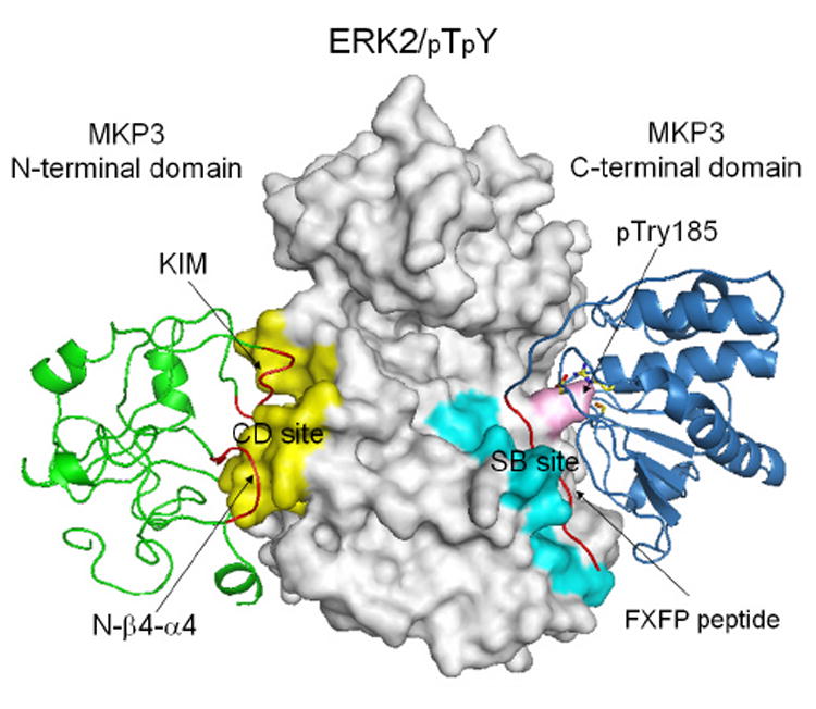
Figure 4.

A structural model for the ERK2/pTpY-MKP3 complex. ERK2/pTpY was shown in gray, MKP3 N-terminal domain in green, and MKP3 C-terminal domain in blue. The common docking (CD) and substrate-binding (SB) site were colored yellow and cyan, respectively, and pTyr185 was colored pink. The kinase interaction motif (KIM) peptide, the N-β4-α4 loop, and the FXFP peptide were highlighted in red. MKP3 active site residues Asp262, Cys293, and Arg299 were also depicted as stick model in atomic colors.
