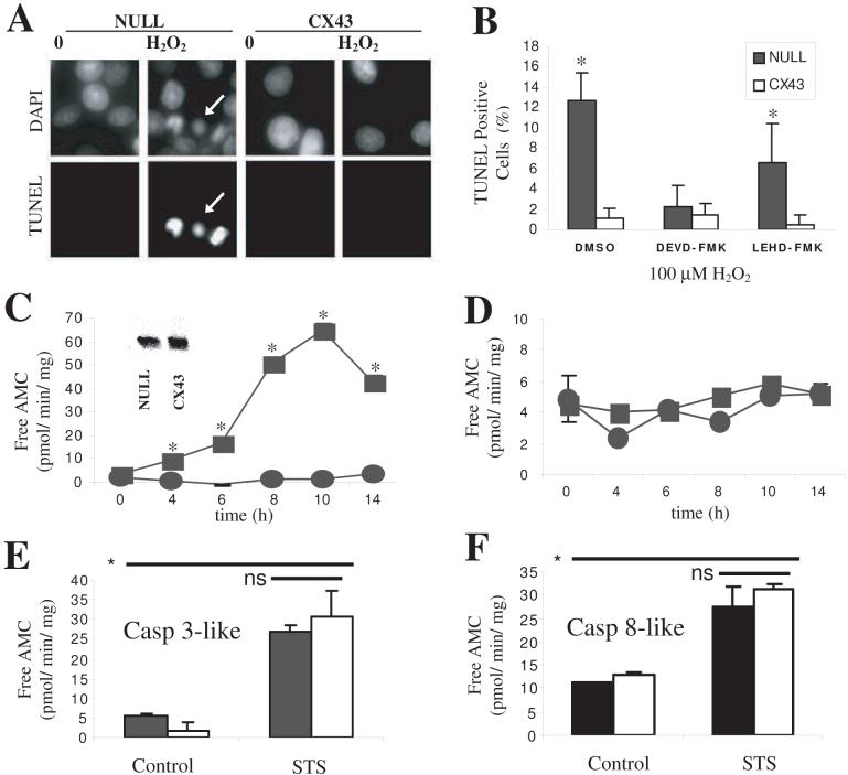
Figure 1. Expression of Cx43 confers relative protection against H2O2-induced apoptosis.
A. C6-null and -Cx cells were exposed to 100μM H2O2 for 15 min and left in serum-free media for an additional 24 h. After fixation, cells were TUNEL labeled and co-stained with DAPI. TUNEL positive cells are clearly seen in C6-null cultures treated with H2O2 (arrows), but not in C6-Cx cells. B. TUNEL labeling was quantified by counting cells from random fields. Where used, caspase inhibitors were pre-incubated for 30 min prior to H2O2 insult and left throughout the duration of the experiment. Greater than 400 cells, from n = 6 experiments were counted. Lysates were prepared from H2O2-injured cells harvested at the indicated times, and proteolysis of (C) Ac-DEVD-AMC (caspase 3-like reporter) or (D) Ac-IETD-AMC (caspase 8-like reporter) was kinetically recorded at 37 C° at 1 min intervals for 1h. Data represents mean ± SD obtained from 3 separate experiments and * indicates significant difference at p<0.05. Inset in C is a Western blot probed with anti-caspase 3 antibody demonstrating equivalent amounts of the caspase protein present in both cell lines. Lysates from STS (1 μM for 6 h) treated cells demonstrated a marked increase in both (E) caspase-3 like and (F), caspase 8-like activity for both cell lines. The means were not significantly different between the C6-null and C6-Cx cell lines under control or STS stimulation. In contrast, STS stimulation significantly increased both caspase-like activities over control.

