
Fig. 7. Monitoring AP dynamics during the course of experiment.
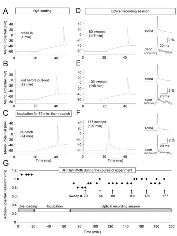
(A – F) Direct current injection (120 pA, 50 ms, standard pulse) was used to trigger somatic AP in different phases of the voltage-imaging experiment. Insets (D-F): simultaneous somatic (whole-cell) and dendrite or axonal (VSD) recording of current-evoked single AP. (G) Plot of AP half-width versus time during the entire course of experiment. APs were probed by standard current pulse during the dye injection phase (Dye loading) and during voltage-imaging (Optical recording session). Patch pipette was not on the cell during Incubation.