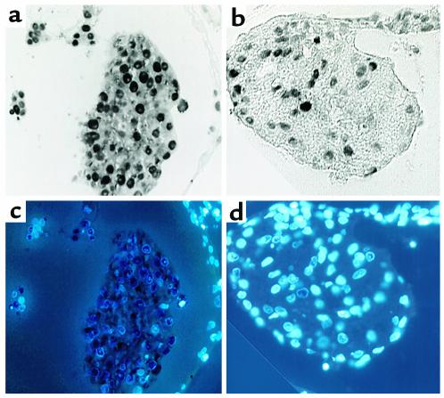
Figure 5.
Ventral pharynx produces a factor that enhances myocardial proliferation. PCNA staining shows proliferating myocytes in strips derived from a single heart cultured in the presence of ventral pharynx (a) or in the presence of ventral pharynx and anti–FGF-neutralizing Ab (b). DAPI staining labeled nuclei in each myocyte allowing determination of the percentage of myocytes that were proliferating in the presence of ventral pharynx (c) or ventral pharynx and anti–FGF-neutralizing Ab (d).

