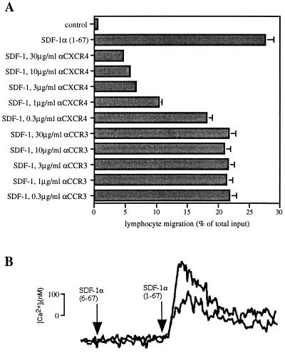
Figure 1.
The CXCR4-specific mAb 12G5 partially blocks SDF-1-induced cellular responses. (A) mAb 12G5 blocks the majority of SDF-1-induced lymphocyte chemotaxis. Freshly isolated PBMC were preincubated with the indicated concentrations of mAbs to CXCR4 (12G5) and CCR3 (7B11) for 15 min and subsequently added to the top chamber of bare filter Transwell inserts. SDF-1 at the optimal concentration of 1.5 μg/ml was added to the bottom chamber. Transmigrated cells were counted by flow cytometry scatter-gating on the lymphocytes. Results are shown as percentage of input. The experiment shown was representative of four independent experiments. (B) 12G5 partially blocks SDF-1-induced increases in intracellular calcium. CXCR4 stably transfected CHO cells (1C2) loaded with fura-2 AM were exposed to 1 μg/ml of full-length SDF-1α-(1–67) or truncated inactive SDF-1α-(6–67) (negative control) in the presence (lower reading) or absence (upper reading) of 10 μg/ml 12G5 mAb. Using the mAb at 50 μg/ml produced identical results.

