Figure 1.
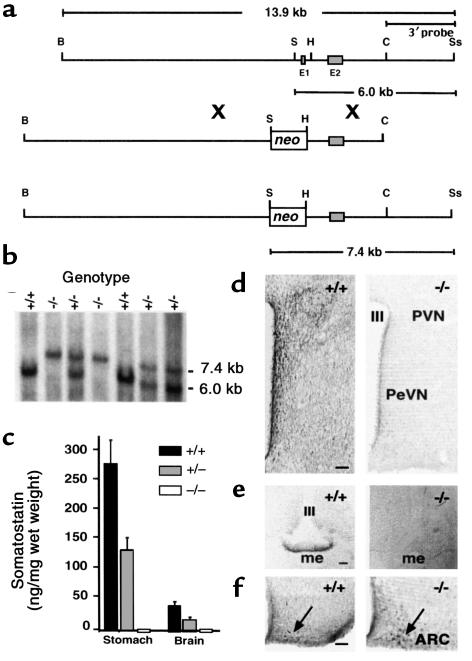
Generation of SST-deficient mice by homologous recombination. (a) Restriction maps of the mouse Smst gene locus (top), targeting vector (middle), and predicted Smst null allele after homologous recombination (bottom) indicate the replacement of exon 1 (E1) and the Smst promoter by a PGK-neomycin selection cassette (neo). B, BamHI; S, SalI; H, HindIII; C, ClaI; Ss, SspI. (b) A Southern blot of genomic DNA digested with SalI and SspI was hybridized with the 3′ probe indicated in a. The 6.0-kb band represents the wild-type allele and the 7.4-kb band represents the Smst null allele in the offspring of an Smst+/– mating pair. (c) RIA of SST in extracts of stomach and total brain (n = 3) demonstrates a 50% reduction in Smst+/– mice and levels below the assay sensitivity in Smst–/– mice. (d) Hemicoronal hypothalamic sections at the level of the periventricular nucleus (PeVN) and anterior paraventricular nucleus (PVN) of an Smst+/+ and an Smst–/– male mouse were immunostained for SST. There is a dense network of immunopositive perikarya and fibers adjacent to the third ventricle (III) only in the Smst+/+ mouse. These signals were also absent after preadsorption of the antiserum with synthetic SST (data not shown). Bars = 100 μm. (e) The median eminence (me) of an Smst+/+ and an Smst –/– mouse were immunostained for SST prohormone. There is no detectable immunoreactivity in the external zone of the me in the Smst–/– mouse. (f) The arcuate nucleus (Arc) of an Smst+/+ and an Smst–/– male mouse immunostained for GHRH. Immunoreactive neuronal perikarya are indicated by the arrows. No difference is apparent between genotypes.

