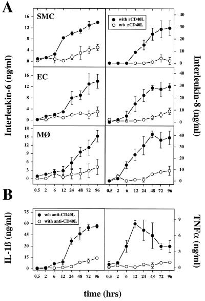
Figure 3.
Induction of proinflammatory cytokine expression in human vascular SMC, EC, or human macrophages by human rCD40L. (A) Supernatants of rCD40L-stimulated (5 μg/ml; •) or unstimulated (○) human vascular SMC, EC, or human macrophages (MØ) were analyzed for IL-6 and IL-8 by ELISA. (B) Macrophages were incubated with rCD40L (5 μg/ml) in the absence (•) or presence (○) of anti-CD40L mAb and analyzed for IL-1β and TNF-α release. Error bars represent SD. Human rCD40L significantly increased cytokine expression relative to control [without (w/o) rCD40L or with anti-CD40L, respectively] after 24 h (P ≤ 0.05, Student’s unpaired t test). Results shown were reproduced for each of the cytokines in three independent experiments.

