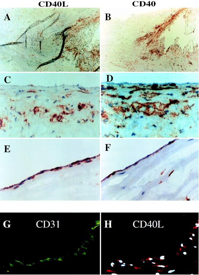
Figure 4.
Expression of CD40L and CD40 in human vascular atherosclerotic lesions in situ. (A and B) Low power views (×40) of frozen sections of human carotid lesions show expression of CD40L and CD40 in the shoulder region of the plaque. (C–F) High power views (×400) of this region revealed specific staining for CD40L and CD40 on SMC and macrophages (C and D), as well as on EC of the luminal border (E and F). As demonstrated here for CD40L on EC (G and H), cell types were characterized by immunofluorescence-double staining as described. The lumen of the artery is at the top of each photomicrograph. Analysis of eight atheroma showed similar results.

