
Figure 3. Staining of embryos in X-gal solution reveals the level and tissue-specificity of MHC-lacZ transgene expression.
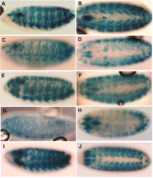
Lateral (left panels) and dorsal (right panels) views are shown. (A, B) Embryos containing pπMHC-lacZ Δi1, which lacks most of intron 1, express ß-galactosidase in body wall and pharyngeal muscles (Ph) in a similar manner to those expressing the minimal myosin promoter pπMHC-lacZ 2 (Fig. 2E). (C, D) Some pπMHC-lacZ Δi1 embryos show differential muscle staining. (E, F) Embryos expressing pMHC1.5-hsplacZ, which contains the intron 1 enhancer, show body wall and pharyngeal muscle staining similar to those expressing pπMHC-lacZ 2. (G, H) Deletion of the 330 terminal bp of pMHC1.5-hsplacZ to yield pMHC1.2-hsplacZ results in greatly reduced body wall muscle expression. (I, J) Expression of pπMHC-lacZ ΔAT, which was produced by deletion of the 330 bp intron fragment from pπMHC-lacZ 2, allows high levels of embryonic ß-galactosidase expression.