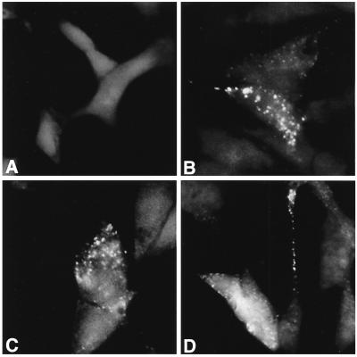
Figure 3.
Analysis of insulin distribution by immunocytochemistry with anti-insulin antibody of transfected clones. (A) Vector-transfected clone V9. (B–D) Calbindin-D28k-transfected clone C2. Shown in B–D are C2 cells from the same batch and passage number. Immunocytochemical staining for insulin was similar to what has been occasionally seen in some similarly fixed and stained RIN cell preparations (M. German, personal communication).

