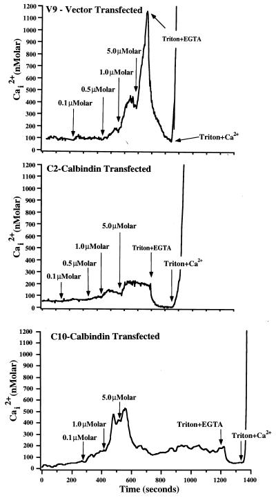
Figure 4.
Representative recordings of the effects of calcium ionophore bromo-A23187 on intracellular Ca2+ in clones transfected with vector (clone V9) or calbindin-D28k (clones C2 and C10). Intracellular Ca2+ was determined in individual cells by microfluorometry of fura-2. The recordings above are the means calculated from 40–45 cells within one field monitored during the course of the experiment. At the times indicated, cells were exposed sequentially to increasing concentrations of bromo-A23187. At the end of each experiment, cells were exposed to Ca2+-free conditions (0.02% Triton X-100 + 10 mM EGTA) and subsequently to high Ca2+ conditions (0.02% Triton X-100 + 10 mM Ca2+) to calibrate dye fluorescence for low and high Ca2+ concentrations. Similar results were observed in 3–4 additional experiments performed with each of these cell clones.

