
Fig. 1.
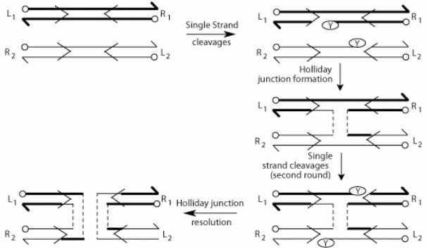
The tyrosine family recombination pathway. The cleavage/exchange of the first and second pair of strands are schematically drawn (details in text). The recombinase binding elements flanking the strand exchange region (spacer) are indicated by the horizontal arrows. The left-right orientation of the recombination partners is designated by L-R. The circular knobs and split arrow heads indicate 5' and 3'ends, respectively.