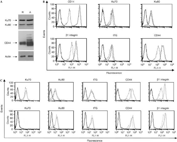
Figure 2.

Ku expression on the cell surface is resistant to inhibitors of the endoplasmic reticulum/Golgi secretory pathway. (A) Whole-cell extracts of resting (R) or activated (A) monocytes were analysed by western blot using the indicated antibodies. (B) Monocytes were incubated in complete medium containing M-CSF in the presence or absence of cycloheximide for 18 h. Cell-surface expression of the indicated antigens in activated monocytes treated (dashed) or not treated (grey) with cycloheximide was analysed by flow cytometry. (C) Monocytes were grown for 18 h in complete medium containing M-CSF in the presence or absence of 5 μg/ml BFA (upper panel) or 20 μM monensin (lower panel). After treatment, cell-surface expression of the indicated antigens was analysed by flow cytometry in treated (dashed) or untreated (grey) monocytes. BFA, brefeldin A; FL1-H, fluorescence pulse 1-height; M-CSF, monocyte colony-stimulating factor; tTG, tissue transglutaminase.
