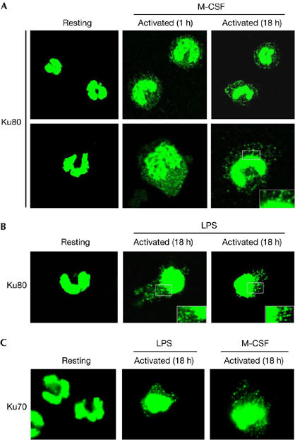
Figure 3.

Ku is translocated from the nucleus to the periphery in monocytes treated with monocyte colony-stimulating factor or lipopolysaccharide. Freshly isolated monocytes or monocytes incubated in complete medium containing M-CSF (A) or LPS (B) for the indicated time period were fixed, stained with the Ku80 (S10B1) antibody and analysed by confocal microscopy. Insets show twofold magnified views of peripheral nuclear regions. (C) Similar experiments were carried out with the Ku70 (S5C1) antibody in resting or activated monocytes as indicated. LPS, lipopolysaccharide; M-CSF, monocyte colony-stimulating factor.
