
Figure 3.
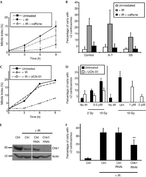
Dependence of DNA-damage-induced centrosome amplification in human cells on ATM/ATR and Chk1 kinase activity. (A) Repression of the G2–M checkpoint by caffeine treatment of human lymphoblastoid cells. Cumulative mitotic indices of human lymphoblastoid cells grown in the presence of colcemid for the time periods indicated after no treatment or after 10 Gy γ-irradiation (IR), with or without preincubation with 2 mM caffeine, as shown. A total of 200 cells were counted per time point in three separate experiments and data presented are the means±s.d. of these replicates. (B) Quantification of control, A-T and Seckel syndrome (SS) cells with multiple centrosomes before and 48 h after 10 Gy ionizing radiation in the presence or absence of caffeine. Centrosomes were counted by immunofluorescence microscopy of γ-tubulin. Data were obtained from at least 500 cells per experiment and histograms show the mean+s.d. of results from three separate blind experiments. (C) UCN-01-mediated abrogation of radiation-induced cell-cycle arrest. Cumulative mitotic indices of human lymphoblastoid cells grown in the presence of colcemid for the time points indicated after no treatment or after 2 Gy γ-irradiation, with or without preincubation with 0.5 μM UCN-01. A total of 200 cells were counted per time point. (D) UCN-01-mediated suppression of centrosome amplification 48 h after the indicated dose of γ-irradiation in the presence or absence of the indicated concentration of UCN-01. Centrosomes were counted by immunofluorescence microscopy of γ-tubulin. Data were obtained from at least 500 cells per experiment and histograms show the mean+s.d. of results from three separate blind experiments. (E) Immunoblot analysis of Chk1 repression in U2OS cells by RNAi at 72 h after transfection, the time at which centrosome counts were performed. Immunoblot for actin was used as a loading control. (F) Chk1 RNA interference-mediated suppression of centrosome amplification 48 h after 20 Gy irradiation of U2OS cells. Centrosomes were counted by immunofluorescence microscopy of γ-tubulin. Data were obtained from at least 500 cells per experiment. Asterisks indicate significant difference from irradiated controls (paired t-test, P<0.01). ATM, ataxia telangiectasia, mutated; ATR, ATM and Rad3-related; Chk1, checkpoint kinase 1; RNAi, RNA-mediated interference.