Figure 4.
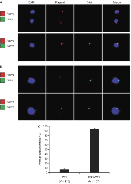
Nuclear positioning of two var loci in different activation states. Nuclei are stained with DAPI (blue). (A) RNA-FISH. Upper panel: nuclear positioning of messenger RNA actively transcribed from the luciferase gene (red) on the plasmid pVLH/IDH-FP in cells in which the chromosomal bsd cassette is silent. Lower panel: nuclear positioning of mRNA of both luciferase (red) and the endogenous bsd cassette (green) when they are both actively expressed. (B) DNA-FISH. Upper panel: nuclear positioning of actively transcribed pVLH/IDH-FP episomes (red) and of a silent bsd cassette under the control of a chromosomal var promoter (green). Lower panel: nuclear positioning when both luciferase (red) carried on the pVLH/IDH-FP episomes and the endogenous bsd cassette (green) are actively expressed. (C) Quantification of colocalization shown in (B). Error bars show standard deviations of multiple, independent counts. DAPI, 4',6-diamidino-2-phenylindole; FISH, fluorescent in situ hybridization; WR, WR99210 selection.

