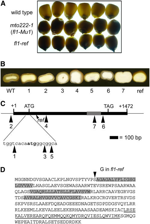
Figure 1.
Phenotype and Gene Structure of the fl1-Mu and fl1-ref Mutations.
(A) Light transmission by mature kernels. Kernels were randomly selected from six ears of W64A+ (top), W64A fl1-Mu1 (mto222-1) (middle), and W64A fl1-ref (bottom) and viewed on a light box.
(B) Kernel phenotypes of the wild type, fl1-Mu1 through fl1-Mu7 (1 to 7), and fl1-ref (ref). Kernel crowns were ground to reveal the thickness of the vitreous endosperm layer.
(C) Diagram of the Fl1 gene. The solid box shows the coding region, and numbered large arrowheads indicate position of Mu insertion alleles. The gray box shows the relative region coding for DUF593. The small arrowhead shows relative position of the fl1-ref point mutation (ref).
(D) FL1 amino acid sequence showing the predicted transmembrane regions (gray boxes) and the conserved DUF593 domain (underlined). The Ser-to-Gly amino acid substitution in fl1-ref is marked with an arrowhead.

