Figure 3.
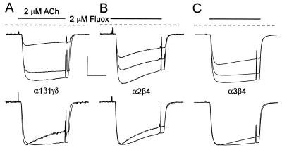
Fluoxetine blockage of AcCho currents mediated by muscle or neuronal nAcChoRs. (A Upper) Superimposed records of AcCho currents from an oocyte expressing muscle α1β1γδ nAcChoRs. The largest trace shows the control current elicited by AcCho alone. After 5–8 min the oocyte was preincubated for 2 min with 2 μM fluoxetine that was maintained during the next AcCho application, which resulted in an inhibited current (record with the lowest amplitude). The middle records show partial recovery after 7–10 min. (A Lower) Normalized and superimposed records corresponding to the same control and blocked AcCho currents. (B and C) Block of AcCho currents by fluoxetine in oocytes expressing neuronal α2β4 and α3β4 nAcChoRs, respectively, following the same protocol as for A. [Horizontal calibration bar = 50 s; vertical calibration bar = 66 nA (A), 800 nA (B), and 400 nA (C).]

