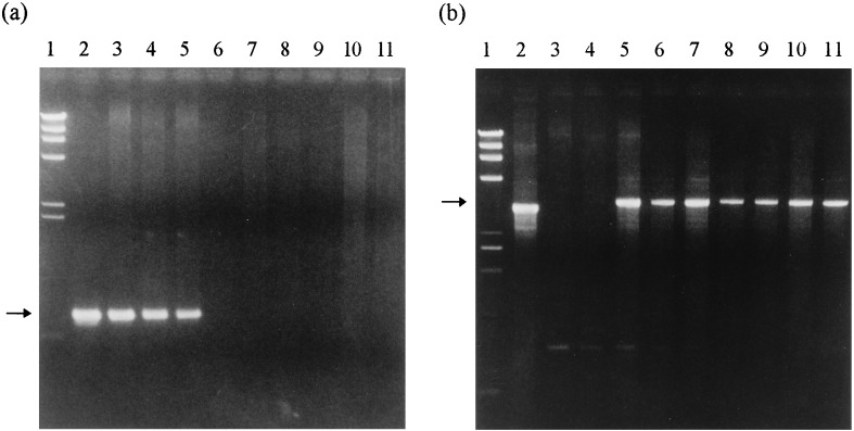
Figure 3.
PCR analysis of ESP shoots and normal plants obtained from ESP shoots. (a) Amplification of the DNA fragments using primers c and d flanking the ipt gene (Fig. 1). The arrow indicates a fragment of approximately 800 bp. (b) Amplification of the fragments using primers a and b (Fig. 1) flanking the position of hit and run cassette. The arrow indicates amplified fragments of 3 kb that result from empty donor sites. Lanes: 1, HindIII size marker (TAKARA shuzo); 2, plasmid pNPI106 in a and plasmid pBI121 in b; 3 and 4, DNA from two independent ESP shoot-derived clones, in which normal shoots did not reappear; 5, DNA from a ESP shoot-derived clone, in which normal shoots reappeared; 6–11, DNA from two independent normal shoots from each of three ESP shoot-derived clones. Lanes 6 and 7 represent 2 shoots from line 2, lanes 8 and 9 represent 2 shoots from line 3, and lanes 10 and 11 represent 2 shoots from line 4.

