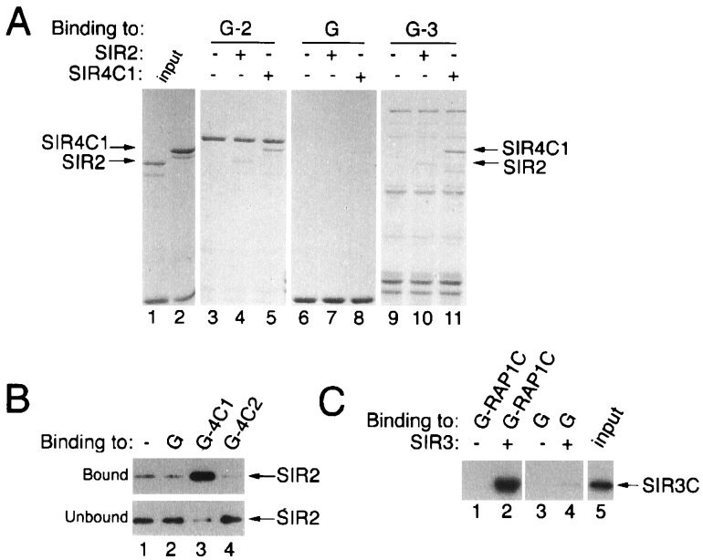
Figure 5.
Direct physical association of bacterially produced SIR2, SIR3, and SIR4 proteins. (A) Coomassie brilliant blue-stained SDS/polyacrylamide gel showing the binding of SIR2 and SIR4C1 (lanes 1 and 2) to GST-SIR2 (G-2, lanes 3–5) and GST-SIR3 (G-3, lanes 9 and 10) proteins. (B) Specific association of SIR2 with GST-SIR4C1 (G-4C1), but not with GST-SIR4C2 (G-4C2), detected by Western blotting with anti-SIR2. (C) Specific association of the C-terminal half of SIR3 (SIR3C) with a GST-RAP1 (G-RAP1C) fusion protein detected by Western blotting with anti-SIR3.

