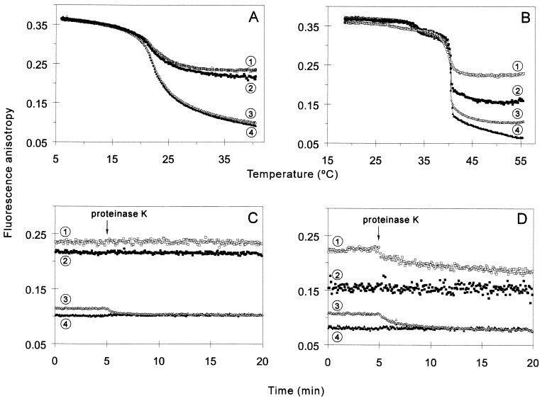
Figure 5.
Effect of GroEL on the membrane physical state tested on LUVs. (A and B) Steady-state fluorescence anisotropy of TMA-DPH (squares, curves 1 and 2) or DPH (triangles, curves 3 and 4) embedded in LUVs made of DMPC (A) or DPPC (B) was measured as a function of temperature in the presence (open symbols, curves 1 and 3) or in the absence (filled symbols, curves 2 and 4) of 1.5 μM GroEL in buffer A. (C and D) Effect of proteinase K treatment on the fluorescence anisotropy of TMA-DPH (squares, curves 1 and 2) or DPH (triangles, curves 3 and 4) in the presence (open symbols, curves 1 and 3) or in the absence (filled symbols, curves 2 and 4) of 1.5 μM GroEL preincubated with 50 μM LUVs of DMPC (C) or DPPC (D), at 35.6°C and 49°C, respectively. Proteinase K (30 μg/ml, final concentration) was added at the indicated time.

