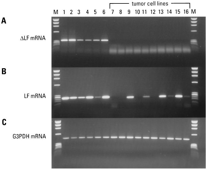
Figure 5.
Tissue expression pattern of ΔLF and LF mRNA in normal compared with tumor-derived cell lines. cDNAs prepared from both normal and tumor cells were used as templates for PCR with the same primer combinations used in Fig. 3. (A) Pattern of ΔLF mRNA in 6 normal tissues (36 cycles of PCR). The spleen and kidney cDNAs were diluted 20× and 10×, respectively. The 10 tumor-derived cells are indicated (42 cycles of PCR). (B) Pattern of LF mRNA in 6 normal tissues (33 cycles of PCR) and 10 tumor-derived cells (42 cycles of PCR). (C) Housekeeping gene control (22 cycles of PCR). Normal tissues: breast (lane 1), spleen (lane 2), placenta (lane 3), testis (lane 4), brain (lane 5), and kidney (lane 6). Tumor cell lines: HeLa cell S3 (lane 7), melanoma A2058 (lane 8), promyelocytic leukemia HL-60 (lane 9), T lymphoblastic leukemia MOLT-4 (lane 10), T lymphoblastic leukemia Jurkat (lane 11), lung tumor A549 (lane 12), breast tumor T41D (lane 13), lymphoma Burkitt’s Raji (lane 14), colorectal adenocarcinoma SW490 (lane 15), and erythroleukemia K562 (lane 16). DNA size markers (φX174 digested with HaeIII) are shown in lane M.

