
Figure 3.
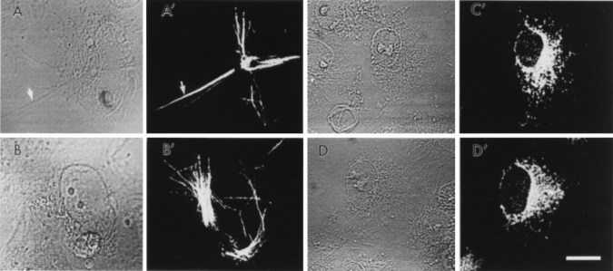
Immunofluorescence microscopy of COS-7 cells transfected with either human FAAH (A and B, cell phase; A′ and B′, anti-FAAH antibody staining) or rat FAAH (C and D, cell phase; C′ and D′, anti-FAAH antibody staining). Note the fibrous, rod-like staining pattern of the human FAAH (arrow) and, in contrast, the more diffuse staining pattern of the rat FAAH. No significant immunostaining was observed in untransfected COS-7 cells, and both the rat and the human FAAH immunoreactivities were effectively competed away by excess peptide antigen.