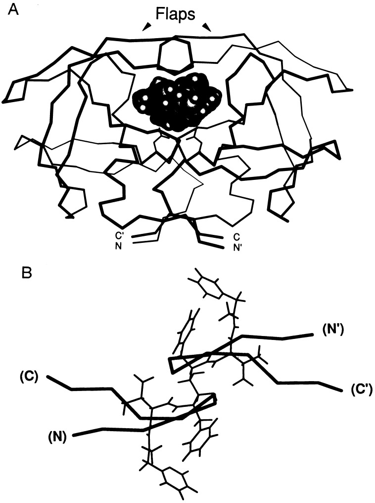
Figure 1.
Flap region of the HIV-1 PR. (A) The α carbon tracing of the HIV-1 PR is shown with the positions of the flaps indicated. The active site aspartic acid residues are shown in wireframe under the bound inhibitor (shown in space filling), and the N and C termini of the two chains of the dimer PR are shown, with the prime (′) used to indicate one of the two chains. This figure was generated using the coordinates of the structure of the HIV-1 PR bound to the inhibitor A-77003 (57). (B) The HIV-1 flap regions in thick line overlaying the inhibitor A-77003 (57). The chain orientation is shown in parentheses. Structures were evaluated using macimdad.

