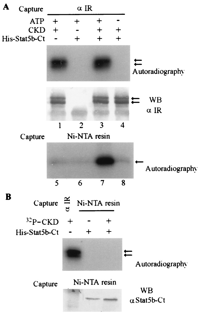
Figure 2.
In vitro phosphorylation assay of His-Stat5b-Ct. (A) His-Stat5b-Ct can be phosphorylated by CKD in vitro. First 0.3 μg of CKD and 0.75 μg of His-Stat5b-Ct proteins were incubated in 50 μl of kinase buffer (10 mM MnCl2/50 mM Tris·HCl, pH 8.0). [γ-32P]ATP (10 μCi) was added to start the reaction. At the end of reaction, 0.5 ml of RIPA buffer was added, and CKD protein was first immunoprecipitated by anti-IR. (Upper) Kinase assay. (Lower) Amount of CKD protein immunoprecipitated and detected by Western blotting with anti-IR antibody. His-Stat5b-Ct protein subsequently was captured from the supernatant by Ni-NTA-resin, washed with 6 M guanidine hydrochloride, followed by water, and then eluted with Laemmli buffer. The phosphorylated His-Stat5b-Ct was detected by autoradiography. (B) His-Stat5b-Ct does not coprecipitate with CKD in vitro. His-Stat5b-Ct was incubated with preautophosphorylated CKD in RIPA buffer and precipitated with Ni-NTA-resin as above. (Upper) Autoradiography. The left lane is the positive control for 32P-CKD precipitated with anti-IR Ab. (Lower) His-Stat5b-Ct protein captured by Ni-NTA-resin and detected by Western blotting with anti-Stat5b-Ct Ab.

