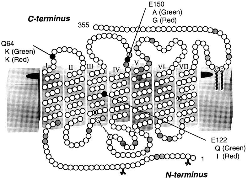
Figure 1.
Amino acid positions in which the electrical properties between the residues chicken rhodopsin and cone visual pigments differ. The transmembrane topography is based on the model of Hargrave et al. (16). Amino acid positions indicated with white circles are those in which rhodopsin and the four types of cone visual pigments have residues similar in electric properties. The gray or black circles indicate the positions at which some (gray) or almost all (black) of the cone pigments have residues electrically different from those of rhodopsin. The residues of rhodopsin replaced in this study are denoted by single-letter codes and numbered using the bovine rhodopsin numbering system. Corresponding residues of chicken green and red are also denoted.

