
Figure 3.
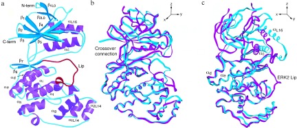
(a) Ribbon diagram of p38 drawn using molscript (38). Helices are purple, β-strands are cyan, and the Lip is red. Note the turn of helix formed by the Lip. (b and c) Superposition of p38 (cyan) and ERK2 (magenta) drawn with setor (39) in two views, approximately 90° apart. Molecules were superimposed using corresponding Cα atoms within the C-terminal domain.