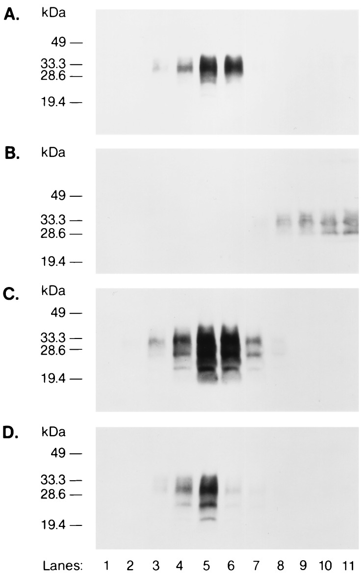
Figure 5.
MHM2-Qa(+) associates with CLDs. (A) ScN2a cells expressing MHM2-Qa(+) were treated with cold Triton X-100 and the CLDs were separated by flotation into sucrose gradients. Gradients were fractionated and fractions tested for presence of Qa molecules by immunoblotting using 3F4 mAb. Lanes 1–7 represent fractions 1–7 of the gradients (5–30% sucrose), lanes 8–11 represent the lysate fractions (40% sucrose). (B) ScN2a cells expressing MHM2-Qa(−) lysed, fractionated and stained as described for A. C and D were equivalent to A and B, respectively, but were stained with α-PrP polyclonal RO73 antiserum, which recognizes endogenous MoPrP. Although the α-PrP polyclonal RO73 antiserum recognizes both MoPrP and MHM2 PrP, only MoPrP is stained in D. Since MHM2-Qa(−) is expressed at levels lower than MoPrP, the staining reaction was terminated before MHM2-Qa(−) was stained by the α-PrP polyclonal RO73 antiserum.

