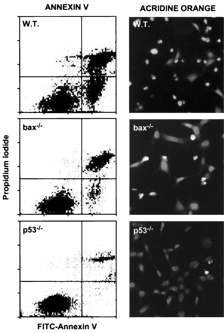
Figure 2.
Apoptosis in bax-deficient E1A-MEFs. E1A-MEFs of the indicated genotype were treated with 0.1 μg/ml adriamycin for 24 h and analyzed for apoptosis by costaining with FITC-annexin V and propidium iodide, or by staining with acridine orange. Annexin V binds phosphotidylserine. Apoptotic changes in membrane biochemistry lead to increased concentration of phosphotidylserine on the outer plasma membrane, where it becomes accessible to annexin V (20). Propidium iodide fluorescently stains late apoptotic cells that have lost membrane integrity. Shown are representative dot plots from two-color flow cytometry: lower left quadrant, viable; lower right quadrant, early apoptotic; upper right, late apoptotic. Acridine orange staining allows visualization of the chromatin condensation characteristic of apoptotic cells.

