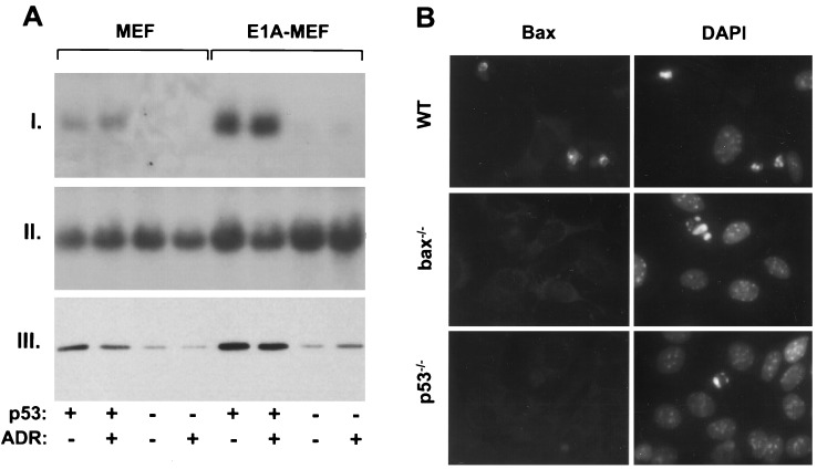
Figure 3.
Endogenous p53 regulates bax expression in MEFs. (A) bax mRNA and protein expression in wild-type and p53−/− MEFs infected with either the E1A-expressing or control virus. Cell populations were treated with 0.1 μg/ml adriamycin for 9 h (ADR) or left untreated. (I) Northern blot using a murine bax cDNA probe (12), or (II) a probe corresponding to the 18S ribosomal RNA. (III) Western blot using an antipeptide rabbit polyclonal primary antibody directed against Bax. (B) Wild-type (WT), bax−/−, and p53−/− E1A-MEFs treated with 0.2 mg/ml adriamycin for 24 h. (Left) Bax expression was measured by immunofluorescence using the Bax antibody described in A. (Right) 4′, 6-diamidono-2-phenylindole (DAPI) staining of the same field reveals the condensed chromatin characteristic of apoptotic cells. The strong immunofluorescent signal observed in wild-type cells was abolished when the Bax antibody was preincubated with the synthetic peptide to which it was generated (not shown).

