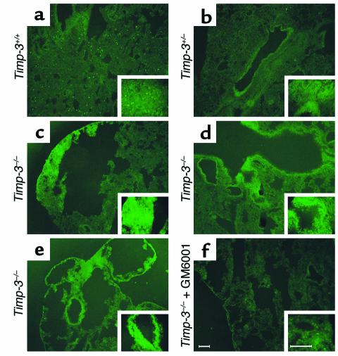
Figure 9.
In situ zymographic analysis of fresh-frozen, noninflated, control and null aged lung. (a) Wild-type lung section demonstrating minimal MMP activity in either the alveolar interstitium or the peribronchiolar regions (upper left corner), represented by cleavage of quenched FITC from a gelatin substrate and subsequent fluorescence. This particular animal appeared to have a higher than usual infiltration of macrophage cells, shown by the focal fluorescence throughout the tissue. Insets show higher magnification. (b) Heterozygotic lung showing slightly elevated MMP activity surrounding a bronchiole. (c) Alveolar interstitium demonstrates heightened MMP activity in the null lung. (d and e) Independent null littermates showing elevated peribronchiolar and interstitial MMP activity. (f) Same animal represented in e, except with the addition of synthetic inhibitor (GM6001) added to the substrate. This negative control demonstrates the majority of the gelatinolytic activity in the null lung is due to MMPs (scale bars, 100 μm).

